During this covid-19 pandemic, chest imaging or chest x-rays are important for the early identification of the coronavirus and to decide the treatment for confirmed and suspected patients with a covid-19 chest infection. These x-rays are fast and cheap. In places where the healthcare centers are far with complex technologies uses these x-rays to identify affected patients. AI in x-rays helps the doctors in a great way for controlling the outbreak of coronavirus. Doctors can measure the severity and can easily classify the disease with the assistance of this chest imaging technology. Artificial Intelligence and cloud computing are used in a digital diagnostic tool that reads vast numbers of X-rays fast and accurately to help doctors prioritize and monitor covid-19 patients. So, doctors can identify the infected people and isolate them thus decreasing the spread of the coronavirus.
Artificial Intelligence Trained With Artificial X-Rays
The speed and accuracy of medical diagnosis can be improved by Artificial Intelligence. But to use AI for identifying the condition seen in X-rays the clinicians have to teach them the algorithm what to look for. For developing AI training sets Prof.Shahrokh came up with a new idea of using machine learning for creating computer-generated X-rays. Certain Simulated X-rays are created that can reflect rare conditions which are then compared with real X-rays providing enough database to train the neural networks. Thus it identifies these conditions in other X-rays. Deep Convolutional Generative Adversarial Network (DCGAN) is an AI technique used for creating these artificial intelligent X-rays. It continuously generates and improves the simulated images.
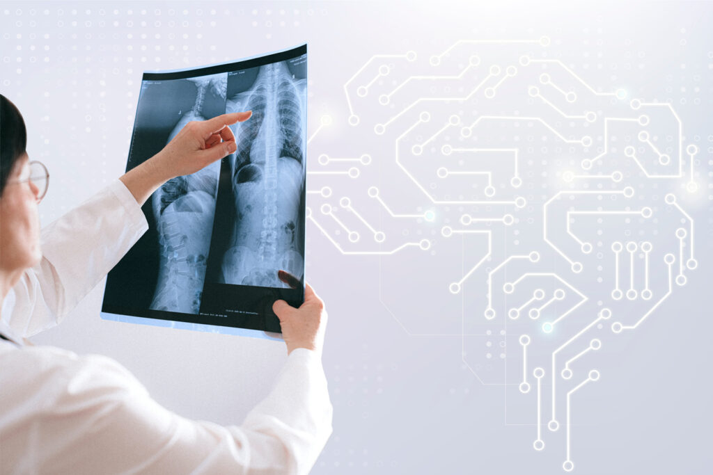
How AI Helps To Analyze Key Findings?
Professor Montana and co-workers developed an AI system that helps to identify key findings by using many adult chest X-rays. For protecting the privacy of patients the images with identifying information are removed. Natural Language Processing is used for pre-processing radiological reports. Each X-ray was labeled with specific abnormalities that are seen on the image because the researchers needed it. NLP uses AI techniques to interpret the framework of each written sentence that helps to identify the clinical findings, body part, and their relationships. Labeling chest X-rays at a range using the NLP system was an important development of professor Montana’s studies.
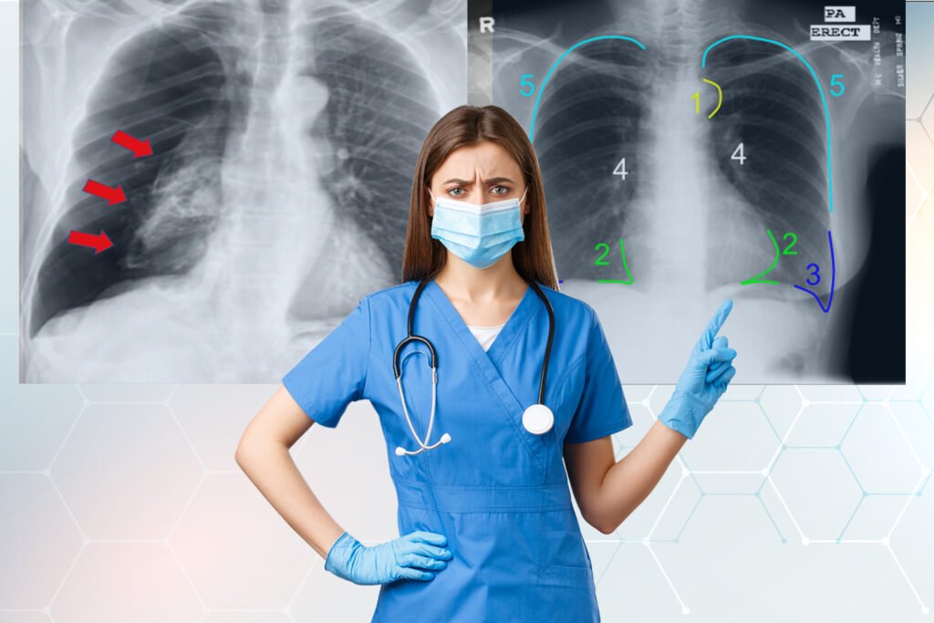
AI Helping Radiologists During Chest X-Ray Review
Dr.Montana is the one who is behind all the research of AI in X-rays that easily differentiate normal from abnormal chest X-rays. AI systems can be well trained using a large database of frequently obtained radiological data and this technology helps to detect all the normal X-rays easily. Thus it reduces the workload of radiologists and provides them more time to focus on critical cases that require more attention. For better performance researchers plan to expand more complex algorithms with a large sample size of research. To assess the performance of prioritized software, a multi-centered study program has been planned in the future. According to Dr. Montana, automated generation of sentences explaining the radiologic abnormalities observed in the images will also be included in the research as a major aspect.
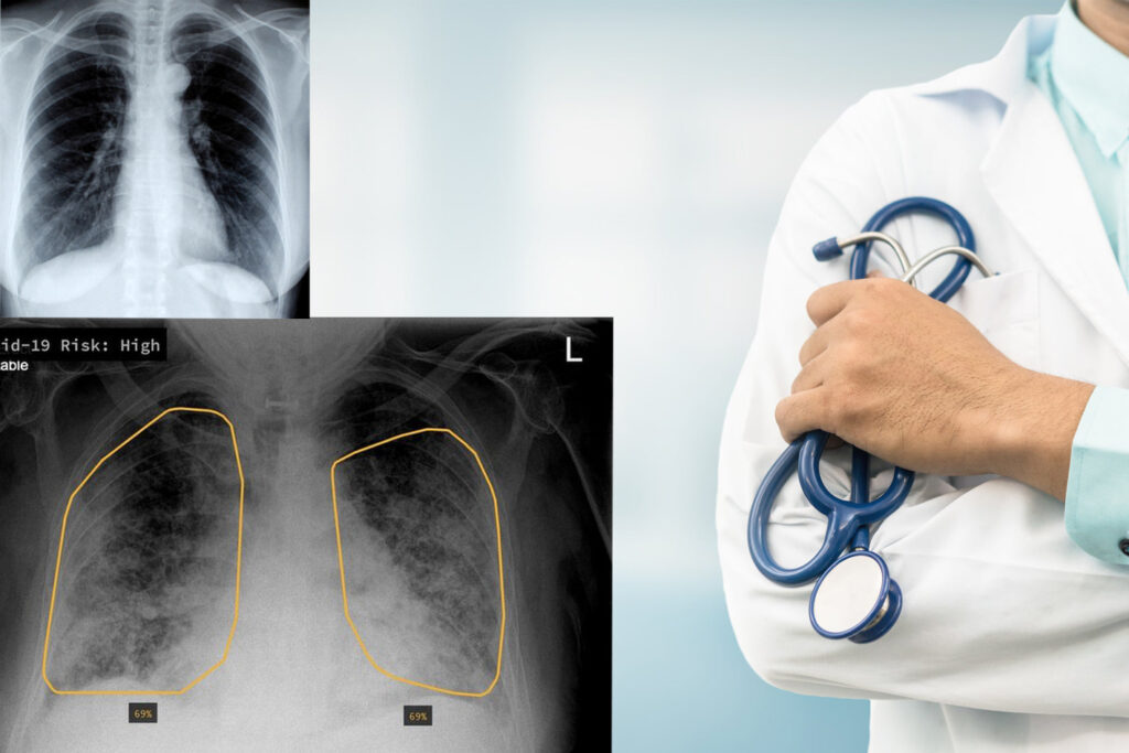
AI Auto-Scanning Chest X-Rays To Identify CoronaVirus
Brunel University in London has developed an AI that automatically identifies coronavirus from lungs X-rays of sick patients. Image analytics and deep learning are used by the algorithm that detects the coronavirus to spot signs and rank them. Targeted X-rays are emphasized by the image processing technique and it also creates visual hints related to certain diseases. The identification and classification algorithm of detecting disease is accurate because it has been tested on real patient’s X-ray images with the confirmed cases. These AI algorithms scan high-quality images and separate the visual cues. It helps in the identification of coronavirus automatically thus helping to monitor the growth of coronavirus in the affected patients.
VisionPro Deep learning
The main goal of visionpro deep learning is the classification of non-covid-19 X-rays from covid-19. A green tool is used by visionpro deep learning for this classification. Images can be trained when it is loaded and labeled once. The Region of Interest(ROI) of a picture can be selected in visionpro deep learning thus making it possible to reduce the edges by 10-20% for removing the borders or letters. In some cases, the edges of images are not removed because many images have lungs towards the edge. So removing these edges may result in the loss of important information. There is no need of resizing images to feed into visionpro deep learning. GUI automatically pre-processes the images of all resolutions and aspect ratios before starting the training. This network training is done by using a high-detail subcategory and by selecting the normal size model.
What We Can Do For You?
AI used in chest X-rays is so helpful to the doctors. They can identify patients who are affected with COVID-19 easily. Identifying and isolating them quickly helps to control the spread of the coronavirus. So because of this great advantage of AI X-rays, most hospitals, clinics, and other screening centers are turning into accurate AI-aided chest X-rays for detecting covid-19 patients. With INFOLKS, you can get perfectly labeled data to train your smart and intelligent medical applications. We have a team of experts solely dedicated to medical data annotation.